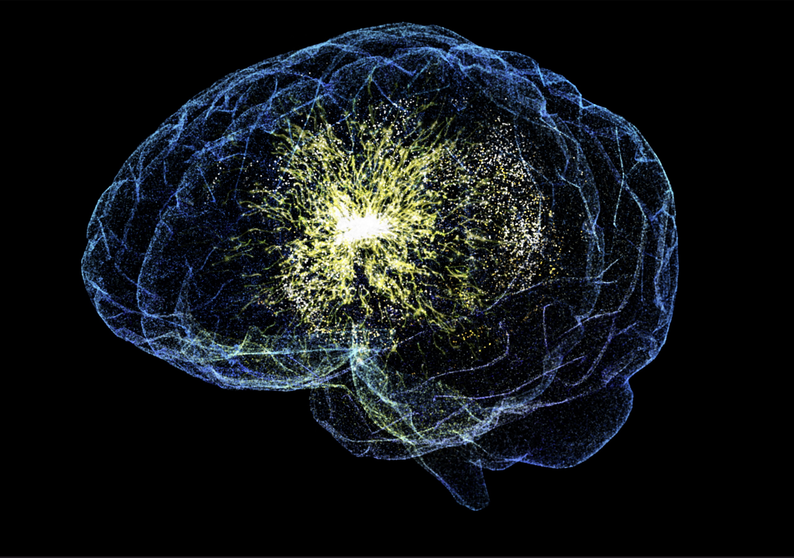
We think of our brains as safe and secure within our skulls, and not easily influenced unless we consume a mind-altering substance, suffer a traumatic injury or undergo invasive brain surgery. However, recent research shows that our brain activity can be influenced non-invasively using nothing but sound and that this technique could have therapeutic potential. As a postdoctoral researcher at UC Berkeley, Dr. Ben Sorum began to think about these types of question while in the Lab of Dr. Stephen G. Brohawn. Now, Dr. Sorum’s current research at Cooper Medical School of Rowan University explores how ultrasound, which can be non-invasively administered from outside the brain and through the skull, can activate specialized proteins in brain cells, changing their activity. The technique, if further developed, may play a key role in the future of neuromodulation, a field with enormous potential for treating neurological disorders. More
For decades, scientists have searched for ways to control neural activity with precision and without causing damage to the brain. Traditional methods such as deep brain stimulation require invasive surgery, while optogenetics, a technique using light to control neurons, requires genetic modification along with the presence of an optical fiber to penetrate and deliver light deep into tissues. Ultrasound, however, presents a compelling alternative: it can penetrate deep into the brain without the need for surgery or implants, offering a potentially revolutionary tool for neuroscience and medicine.
The initial focus was on a specific protein called the TRAAK ion channel that is present in myelinated neural cells. TRAAK is a mechanosensitive potassium channel, meaning that it responds to mechanical forces, such as pressure or stretching within the cell membrane, which open the channel to allow potassium to travel across the membrane. The researchers discovered that ultrasound waves could activate these channels, mimicking the effects of mechanical stimulation. This activation likely occurs because ultrasound increases membrane tension, causing the channel to open and allowing potassium ions to flow, thereby influencing neural activity.
To confirm their findings, the team conducted experiments using different biological models, including purified TRAAK channels embedded in artificial membranes, as well as in samples from mice and frogs. The results were striking: ultrasound was able to activate TRAAK channels in all these models, even in the absence of other cellular components. This suggests that ultrasound does not require intermediary molecules or complex signaling pathways to exert its effect, and it directly influences the lipid membrane to trigger ion channel activity.
Another critical observation was the speed of activation. The TRAAK channels responded to ultrasound stimulation within microseconds, making this a highly efficient method for influencing neural activity. Moreover, the researchers found that varying the intensity and duration of the ultrasound pulses allowed them to finely tune the extent of TRAAK activation. This level of precision is crucial for potential therapeutic applications, as it could enable doctors to target specific brain regions with minimal side effects.
Building on these findings, the researchers expanded their focus to include two additional mechanosensitive potassium ion channels: TREK-1 and TREK-2. These channels, along with TRAAK, belong to a family of ion channels known for their role in sensory processing and muscle contraction. The follow-up study aimed to understand how these channels differ in their sensitivity to membrane tension and how ultrasound could be used to activate them.
Using electrophysiological techniques and advanced imaging, the team measured the forces required to activate TRAAK, TREK-1, and TREK-2. Their findings revealed that TRAAK is the most sensitive to membrane tension, while TREK-1 and TREK-2 require stronger forces to open. This suggests that different channels may serve distinct roles in the nervous system, with TRAAK likely responding to subtle mechanical changes and TREK channels acting under more intense conditions.
The researchers employed a combination of patch-clamp recording, a laboratory technique that measures the ionic currents flowing through a cell or tissue, and membrane imaging to quantify the tension response of these channels. They observed that TRAAK and TREK-1 were activated broadly over a range of physiological tensions, whereas TREK-2 exhibited a sharper, switch-like response similar to mechanosensitive channels found in bacteria. This finding indicates that TRAAK and TREK-1 may be more suited for gradual, graded responses to mechanical stimuli, whereas TREK-2 may function in situations requiring rapid activation.
To confirm their hypothesis, the researchers tested how ultrasound stimulation affects these channels. Their experiments demonstrated that low-intensity, low-frequency ultrasound effectively increased membrane tension, leading to TRAAK activation. Interestingly, this response closely mirrored the effects of direct mechanical pressure, reinforcing the idea that ultrasound can be used as a precise tool for neuromodulation. Furthermore, imaging confirmed that ultrasound-induced changes in membrane curvature coincided with ion channel activation, providing direct evidence of the mechanism at play.
One of the most exciting implications of this research is its potential application in treating neurological conditions. Many disorders, including epilepsy, depression, and chronic pain, involve abnormal neural activity. By harnessing ultrasound to control specific neurons, scientists may be able to develop non-invasive therapies that help to counteract disease processes and restore healthy brain function. Furthermore, because TRAAK and TREK channels are found in nodes of Ranvier, which are the small gaps between myelinated sections of neurons where electrical signals are regenerated, targeting these channels may enhance neural communication in conditions where this is an issue, such as multiple sclerosis or nerve damage.
Additionally, the findings have important implications for pain management. TREK-1 and TREK-2 have been implicated in the body’s pain pathways, suggesting that ultrasound stimulation of these channels could provide a novel method for alleviating chronic pain. By modulating these channels in the peripheral nervous system, researchers could develop ultrasound-based therapies for pain relief without the need for drugs or invasive procedures.
The research also paves the way for ‘sonogenetics’, a technique where specific cells are genetically modified to be more responsive to ultrasound. By engineering neurons to express more TRAAK channels, for instance, scientists could enhance the brain’s sensitivity to ultrasound stimulation, allowing for even greater precision in targeting brain circuits without affecting surrounding tissues. This could be particularly beneficial for treating conditions where more selective neuronal stimulation is needed to maintain or restore function.
The findings mark a significant step forward in our understanding of how ultrasound interacts with the brain at the molecular level. Their combined studies show that ultrasound can be used to activate mechanosensitive ion channels with remarkable precision, offering a new avenue for non-invasive brain stimulation.
As research progresses, this technique could transform the way that we treat neurological disorders, offering a future where brain function can be tuned with the gentle touch of sound waves. If further studies confirm its safety and effectiveness, ultrasound-based neuromodulation could become a game-changing tool in medicine, providing a new method for treating some of the most challenging brain disorders.